All content on this site is intended for healthcare professionals only. By acknowledging this message and accessing the information on this website you are confirming that you are a healthcare professional. If you are a patient or carer, please visit Know AML.
The AML Hub website uses a third-party service provided by Google that dynamically translates web content. Translations are machine generated, so may not be an exact or complete translation, and the AML Hub cannot guarantee the accuracy of translated content. The AML Hub and its employees will not be liable for any direct, indirect, or consequential damages (even if foreseeable) resulting from use of the Google Translate feature. For further support with Google Translate, visit Google Translate Help.


The AML Hub is an independent medical education platform, sponsored by Daiichi Sankyo, Kura Oncology, Syndax, Abbvie, Thermo Fisher and Johnson & Johnson. Funders are allowed no direct influence on our content. The levels of sponsorship listed are reflective of the amount of funding given. View funders.
Now you can support HCPs in making informed decisions for their patients
Your contribution helps us continuously deliver expertly curated content to HCPs worldwide. You will also have the opportunity to make a content suggestion for consideration and receive updates on the impact contributions are making to our content.
Find out more
Create an account to access:
Bookmark & personalize site content
Receive alerts for new content in your areas of interest
View AML content recommended for you
Clinical outcomes and prognostic value of MRD in patients with NPM1-mutated AML
Mutations in nucleophosmin (NPM1) are the most common genetic alterations in acute myeloid leukemia (AML), accounting for 20–30% of adult cases.1 Given its prognostic significance, NPM1-mutated AML is classified as a distinct entity according to the World Health Organization (WHO) and European Leukemia Net (ELN); these mutations often occur in exon 12 of the NPM1 gene, resulting in the cytoplasmic accumulation of NPM1 (termed NPM1c).1
NPM1c regularly coincides with mutations in FMS-like tyrosine kinase 3 (FLT3), isocitrate dehydrogenase (IDH)1 and IDH2, and DNA methyltransferase 3 alpha (DNMT3A).1 Interestingly, NPM1c without FLT3-ITD mutations is associated with favorable prognostic outcomes in patients with newly diagnosed AML; although this impact is strongly dependent on the presence of concurrent mutations. However, little is known about its prognostic impact and therapeutic implications among patients with relapsed/refractory (R/R) AML.1
Treatment regimens including venetoclax-based combinations have significantly improved clinical outcomes in patients with NPM1-mutated AML.2 In addition, molecular measurable residual disease (MRD) assessments are strongly prognostic among patients with NPM1-mutated AML treated with intensive chemotherapy, and MRD by flow cytometry is prognostic across molecular subgroups treated with venetoclax. However, little is known about the prognostic significance of molecular MRD in patients with NPM1-mutated AML treated with these regimens.2
Here, we summarize a study by Issa et al.1 on the prognostic impact and clinical outcomes of patients with NPM1-mutated R/R AML across different lines of salvage therapies. We also summarize an analysis of the prognostic role of MRD in patients with NPM1mutated AML receiving non-intensive venetoclax-based therapies, presented by Othman2 at the 64th American Society of Hematology (ASH) Annual Meeting and Exposition.
Prognostic impact and clinical outcomes of NPM1-mutations in R/R AML1
Study design
This was a retrospective analysis of adult patients with R/R AML treated either with high-intensity (HI) or low-intensity (LI) regimens, depending on age and co-morbidities, between September 2012 and December 2020. HI regimens included cytarabine and idarubicin combinations with or without the addition of a nucleoside analog such as cladribine, fludarabine, or clofarabine. LI regimens included hypomethylating agents (5-azacitidine or decitabine) or low-dose cytarabine, with the addition of venetoclax.
Outcomes included:
- Response rates, defined according to the International Working Group recommendations
- Overall survival (OS), defined as the time from treatment start date in relapsed disease until the time of death or last follow-up
- Relapse free survival (RFS), defined as the time of complete remission (CR)/CR with incomplete count recovery (CRi) until relapse or death
Results
Of the 1,722 patients with R/R AML included in this analysis, 206 had NPM1c and 1,516 had NPM1wt. Baseline characteristics and mutational co-occurrence patterns of NPM1c versus NPM1 wild type (NPM1wt) are summarized in Table 1. The co-occurrence of NPM1c with DNMT3A, FLT3-ITD, IDH1/2, and WT1 was significantly higher compared with NPM1wt. However, co-occurrence of mutations in ASXL1, RUNX1, TET2, and TP53 was lower in NPM1c compared with NPM1wt.
Table 1. Baseline characteristics of patients with NPM1c versus NPM1wt*
|
AML, acute myeloid leukemia; BM, bone marrow; S1, salvage 1; S2, salvage 2; S3, salvage 3; WBC, white blood cell. |
|||
|
Characteristic, % (unless |
NPM1c |
NPM1wt |
p value |
|---|---|---|---|
|
Median age (range), years |
64 (17–91) |
64 (16–90) |
0.6 |
|
Sex |
|
|
|
|
Male |
42 |
62 |
<0.0001 |
|
Female |
58 |
38 |
— |
|
Median hemoglobin (range), g/dL |
9.3 (6–15) |
9.1 (4–18) |
0.1 |
|
Median WBC (range), × 109/L |
8.1 (0.1–227) |
3.6 (0.1–339) |
<0.0001 |
|
Median platelet count (range), × 109/L |
45 (4–624) |
43 (1–1,552) |
0.007 |
|
Median peripheral blast (range) |
37 (0–100) |
8 (0–100) |
<0.0001 |
|
Median BM blast (range) |
60 (0–99) |
30 (0–98) |
<0.0001 |
|
t-AML |
8 |
15 |
0.007 |
|
s-AML |
3 |
16 |
<0.0001 |
|
Cytogenetics† |
|
|
|
|
Diploid |
61 |
31 |
— |
|
Complex, -5, -7 |
8 |
38 |
— |
|
Other |
31 |
31 |
— |
|
Mutations† |
|
|
|
|
DNMT3A |
50 |
17 |
<0.0001 |
|
FLT3-ITD |
49 |
11 |
<0.0001 |
|
TET2 |
31 |
65 |
<0.0001 |
|
IDH1 |
20 |
7 |
<0.0001 |
|
IDH2 |
20 |
11 |
0.0003 |
|
KRAS/NRAS |
19 |
19 |
1.0 |
|
WT1 |
19 |
10 |
0.009 |
|
FLT3-D835 |
13 |
4 |
<0.0001 |
|
ASXL1 |
6 |
22 |
<0.0001 |
|
TP53 |
6 |
25 |
<0.0001 |
|
RUNX1 |
5 |
22 |
<0.0001 |
|
Lines of therapy |
|
|
|
|
S1 |
64 |
63 |
0.8 |
|
S2 |
15 |
18 |
0.3 |
|
≥S3 |
21 |
19 |
0.5 |
|
Duration of first remission‡ |
|
|
|
|
≤6 months |
41 |
40 |
1.0 |
|
6–12 months |
27 |
25 |
0.9 |
|
≥12 months |
32 |
34 |
0.9 |
Mutational landscape in patients with NPM1-mutated AML at relapse
Following first-line treatment, 76% of patients achieved clinical remission. An overall increase in ELN risk was observed among the remaining patients with relapsed disease (24%), with a 10–16% increase in ELN-intermediate risk and 12–25% increase in ELN-adverse risk (p = 0.01, each); thus indicating mutational changes from diagnosis to relapse.
Loss in NPM1c (n = 6) versus persistence of NPM1c (n = 206) at relapse was significantly associated with improved 1-year RFS (80% vs 34%; p = 0.5) and 1-year OS (83% vs 30%; p = 0.002), suggesting NPM1c loss at relapse indicative of a de novo AML.
Among patients with NPM1c, similar CR/CRi rates were observed in patients with or without co-existing FLT3-ITD mutations (55% vs 58%). Improved OS was seen in patients with NPM1c and FLT3wt compared with NPM1c and FLT3-ITD co-mutations (median OS, 8.6 months vs 5.8 months; p = 0.05). Similar response rates were also observed between NPM1c concurrent with DNMT3A, IDH1, TET2, or RAS versus NPM1c and the corresponding wild-type genes. On the other hand, NPM1c and IDH2 comutations were associated with a significantly higher CR rate (50% vs 27%; p = 0.03) and an improved OS (median OS, 14.5 months vs 5.8 months; p = 0.01) compared with NPM1c and IDH2wt.
Clinical outcomes by line and type of therapy
Overall, higher response rates were observed in the NPM1c versus NPM1wt cohort following each line of salvage therapy; though this difference was less noticeable in salvage therapy 2 and 3 (Figure 1).
Figure 1. Response rates (CR/CRi) by line of therapy*
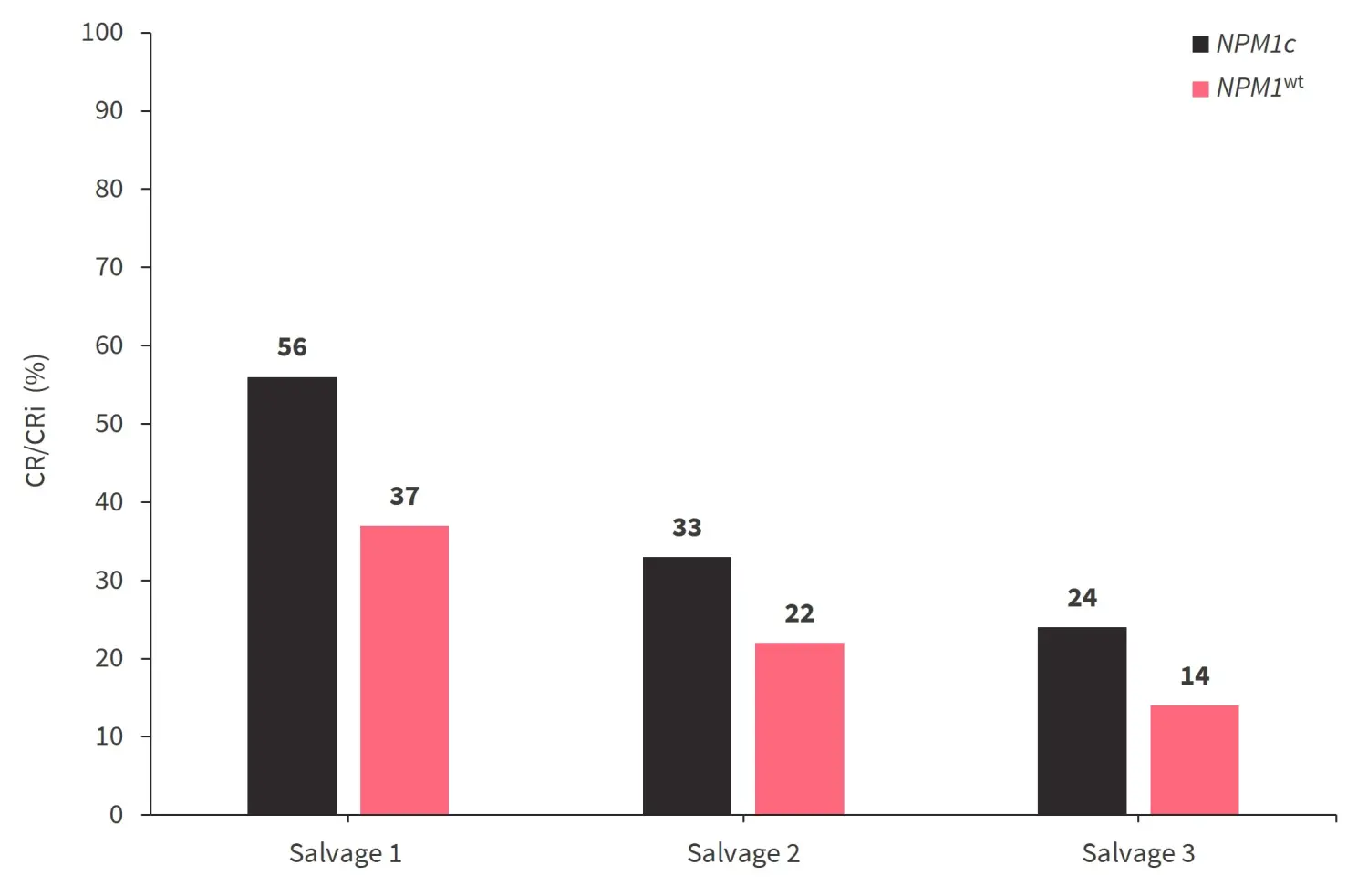
CR, complete response; CRi, CR with incomplete count recovery; NPM1c, cytoplasmic accumulation of NPM1; NPM1wt, NPM1 wild type.
*Adapted from Issa, et al.1
A trend towards an improved OS and RFS was observed among NPM1c versus NPM1wt following the first line of therapy, and similar outcomes were observed between the two cohorts in subsequent lines of therapy (Table 2).
Table 2. Median RFS and OS outcomes*
|
Factor, months |
Median RFS |
Median OS |
||
|---|---|---|---|---|
|
NPM1c |
NPM1wt |
NPM1c |
NPM1wt |
|
|
Overall |
5.5 |
5.6 |
6.1 |
5.5 |
|
By line of therapy |
|
|
|
|
|
S1 |
8.3 |
5.7 |
7.8 |
6.0 |
|
S2 |
3.3 |
5.1 |
5.3 |
4.1 |
|
S3 |
4.0 |
5.4 |
3.5 |
3.6 |
|
By diploid karyotype |
7.6 |
5.5 |
8.0 |
7.9 |
|
By age |
|
|
|
|
|
<60 years |
11.7 |
6.9 |
8.1 |
6.6 |
|
≥60 years |
4.3 |
5.2 |
5.1 |
5.1 |
|
NPM1c, cytoplasmic accumulation of NPM1; NPM1wt, NPM1 wild type; OS, overall survival, RFS, relapse-free survival; S1, salvage 1; S2, salvage 2; S3, salvage 3. |
||||
Response rates were significantly higher in NPM1c versus NPM1wt cohort treated with HI regimens (p < 0.0001). A substantially higher response rate was observed with the addition of venetoclax to LI (p = 0.02) in NPM1c versus NPM1wt cohort (Figure 2), translating into a clinically improved RFS (median RFS, 15.8 months vs 4.6 months; p = 0.05) and OS (median OS, 14.7 months versus 5.9 months, p = 0.02). Among patients with NPM1c, outcomes for LI + venetoclax and HI regimens were matched with a median OS of 14.5 months versus 8.1 months, respectively (p = 0.4).
Figure 2. Response rates (CR/CRi) by type of therapy*
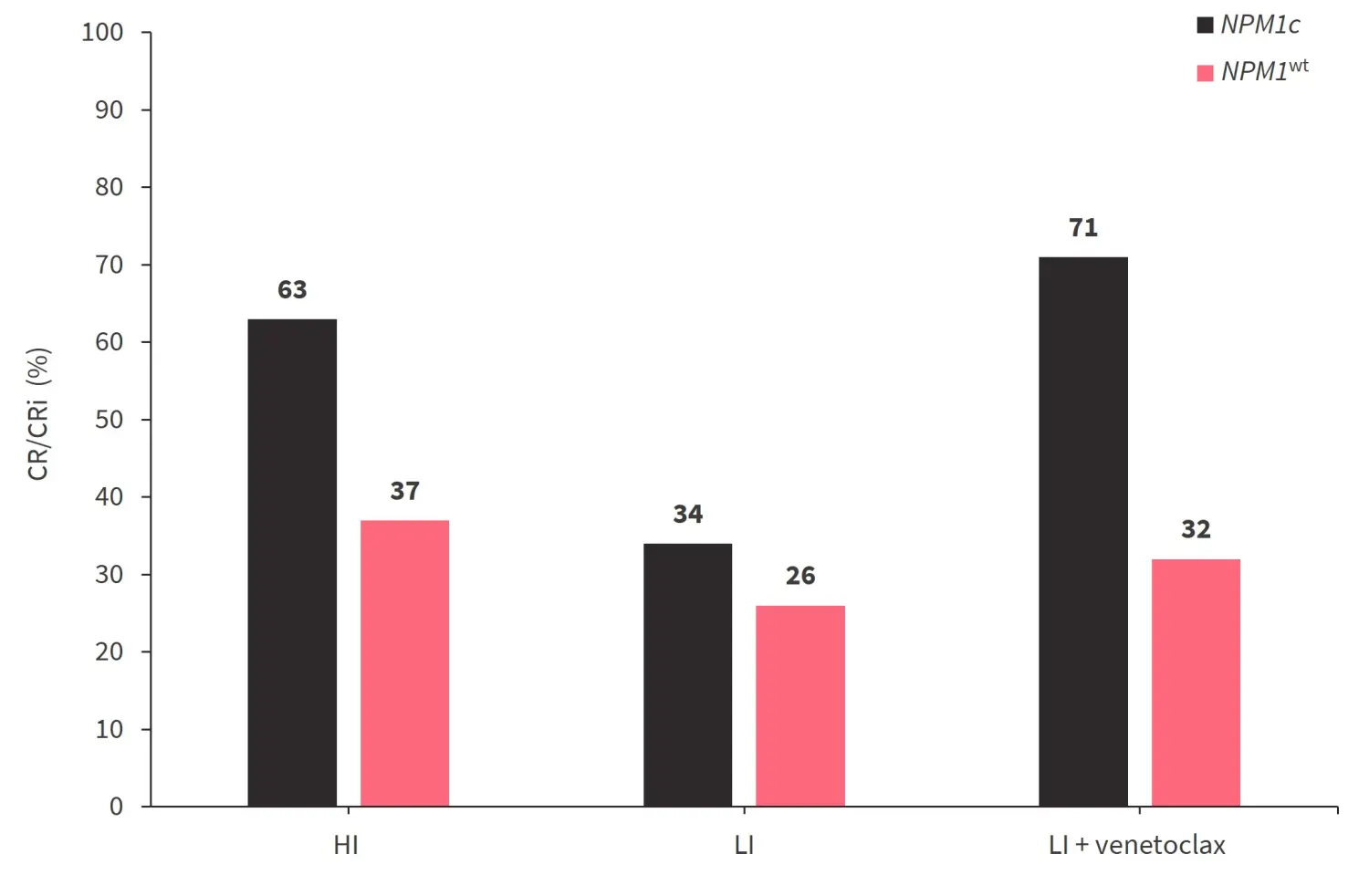
CR, complete response; CRi, CR with incomplete count recovery; HI, high intensity, LI, low intensity; NPM1c, cytoplasmic accumulation of NPM1; NPM1wt, NPM1 wild type.
*Adapted from Issa, et al.1
With regards to NPM1/FLT3 co-mutations, an FLT3 inhibitor either as a monotherapy or in combination yielded a 57% overall response in patients. However, IDH inhibitor-based therapies yielded a response rate of 33% in patients with NPM1c and IDH co-mutations. Patients with NPM1c-mutated AML receiving hematopoietic stem cell transplantation compared with those not receiving hematopoietic stem cell transplantation had an improved RFS (median RFS, 20.7 months vs 4.0 months; p < 0.0001) and OS (22.2 months vs 8.6 months; p < 0.0001), respectively.
In the multivariate analysis, NPM1 mutational status was not prognostic for OS; however older age (≥60 years), duration of first remission <6 months (<0.001) and TP53mut (p = 0.001) were independent prognostic factors associated with worse OS in patients with R/R AML.
Prognostic role of MRD in patients with NPM1mut AML treated with venetoclax2
Study design
This retrospective cohort included 45 patients and 10 patients from a UK and Australian-based real-world AML cohort, respectively. Eligible patients who harbored the NPM1 mutation and were treated with frontline LI venetoclax-based regimens had achieved CR with or without CRi and had at least one bone marrow (BM) MRD assessment in the first 6 months using quantitative polymerase chain reaction.
Results
A total of 55 patients were included in this analysis. Baseline characteristics are highlighted in Table 3.
Table 3. Baseline characteristics*
|
AML, acute myeloid leukemia; ECOG, Eastern Co-operative Oncology Group; LDAC, low-dose cytarabine. |
|
|
Characteristics, % (unless otherwise stated) |
Total cohort |
|---|---|
|
Median age, years |
72.7 |
|
ECOG ≤1 |
87.0 |
|
Type of AML |
|
|
De novo |
81.0 |
|
Secondary |
13.0 |
|
Therapy-related |
5.6 |
|
Cytogenetic risk |
|
|
Intermediate |
91.0 |
|
Adverse |
3.7 |
|
Mutations |
|
|
FLT3-ITD |
30.0 |
|
FLT3-TKD |
15.0 |
|
DNMT3A† |
33.0 |
|
IDH1† |
10.0 |
|
IDH2† |
21.0 |
|
TP53† |
2.4 |
|
Type of treatment received |
|
|
LDAC |
45.0 |
|
Azacitidine |
51.0 |
|
Decitabine |
4.0 |
The median BM MRD assessment within the first 6 months of therapy was 2 (range, 1–4) showing a steady decrease over time, with a median MRD undetectable at 4 months. Median MRD negativity was also attained in peripheral blood (PB) at 3 months The best MRD response within the 6 months was achieved by 46% of patients, with a median time to first MRD undetectable of 97 days, a ≥4 log-fold NPM1 copy reduction in 19%, and a <4 log-fold reduction in 35% of patients.
Rates of MRD negativity by subgroups were as follows: de novo versus secondary disease (50% vs 20%), normal versus abnormal karyotype (48% vs 33%), no ITD versus ITD (53% vs 25%), DNMT3Amut versus DNMT3Awt (43% vs 54%), IDH1mut versus IDH1wt(75% vs 47%), and IDH2mut versus IDHwt (78% vs 42%).
MRD responses and survival outcomes
At a median follow up of 24.3 months, the 2-year OS and molecular EFS within the overall-treated cohort were 61% and 60%, respectively. Patients with a deeper MRD reduction yielded better EFS and OS outcomes, with MRD negativity yielding the best outcome, a ≥ 4-log fold change achieving intermediate outcomes, and <4 log fold reduction yielding poorer outcomes (p = 0.00027 for OS and p < 0.0001 for EFS).
An MRD threshold of <0.005 NPM1 copies per 100 ABL (reference gene) was identified as the best prognosticator of EFS and OS. In the multivariate analysis, age and MRD above copy number threshold were significantly associated with OS (p = 0.039 and p = 0.002, respectively).
A total of 74% of patients with PB samples achieved MRD negativity, while MRD positivity was associated with poor outcomes (p = 0.02). Although, detectable PB MRD is less sensitivity compared with BM, MRD positivity in BM and MRD negativity in PB demonstrated similar OS to MRD negativity in both BM and PB, suggesting the potential of PB to adequately predict outcomes.
Conclusion
The retrospective analyses by Issa et al.1 showed that NPM1-mutated AML has minimal impact on prognostic outcomes in patients with R/R AML. However, the addition of venetoclax to salvage therapies significantly improved the outcomes in this population. Studies investigating novel treatment strategies are needed to further improve outcomes in this molecular subgroup. Results from the retrospective analysis by Othman2 showed that MRD assessments by quantitative polymerase chain reaction were strongly prognostic of clinical outcomes in patients with NPM1-mutated AML treated with LI venetoclax combination therapies. A threshold of <0.005 NPM1 copies per 100 ABL in BM samples was predictive of the high OS and was independent of pretreatment variables in patients with NPM1mut AML.
References
Please indicate your level of agreement with the following statements:
The content was clear and easy to understand
The content addressed the learning objectives
The content was relevant to my practice
I will change my clinical practice as a result of this content
Your opinion matters
What is the typical turnaround time for next-generation sequencing (NGS) results for AML at your center?

