All content on this site is intended for healthcare professionals only. By acknowledging this message and accessing the information on this website you are confirming that you are a healthcare professional. If you are a patient or carer, please visit Know AML.
The AML Hub website uses a third-party service provided by Google that dynamically translates web content. Translations are machine generated, so may not be an exact or complete translation, and the AML Hub cannot guarantee the accuracy of translated content. The AML Hub and its employees will not be liable for any direct, indirect, or consequential damages (even if foreseeable) resulting from use of the Google Translate feature. For further support with Google Translate, visit Google Translate Help.


The AML Hub is an independent medical education platform, sponsored by Daiichi Sankyo, Johnson & Johnson, Syndax, Thermo Fisher Scientific, Kura Oncology, and AbbVie. Funders are allowed no direct influence on our content. The levels of sponsorship listed are reflective of the amount of funding given. View funders.
Now you can support HCPs in making informed decisions for their patients
Your contribution helps us continuously deliver expertly curated content to HCPs worldwide. You will also have the opportunity to make a content suggestion for consideration and receive updates on the impact contributions are making to our content.
Find out more
Create an account to access:
Bookmark & personalize site content
Receive alerts for new content in your areas of interest
View AML content recommended for you
Disparities in survival and treatment outcomes between Black and White AYAs with AML
Adolescents and young adults (AYAs) diagnosed with acute myeloid leukemia (AML) generally demonstrate superior outcomes when compared with older patients (≥40 years old). Disparities have been highlighted between Black and White patients with AML aged 18–60 years, with poorer survival observed in the Black population. Until now, it remained unknown whether these inconsistencies persisted in AYAs with AML.
Karilyn Larkin and colleagues1 sought to determine survival rates, treatment outcomes, and genetic profiles of non-hispanic Black AYAs with AML compared to those observed in the non-hispanic White population. The results from their study were presented at the 63rd American Society of Hematology (ASH) Annual Meeting and Exposition.1
Study design
A total of 655 AYAs were included in this analysis comparing the molecular characteristics and survival outcomes of patients with AML who self-identify as Black (n = 89) or White (n = 566) undergoing treatment with standard intensity cytarabine/anthracycline induction therapy between 1986 and 2016 (Figure 1).
Figure 1. Patient cohort*
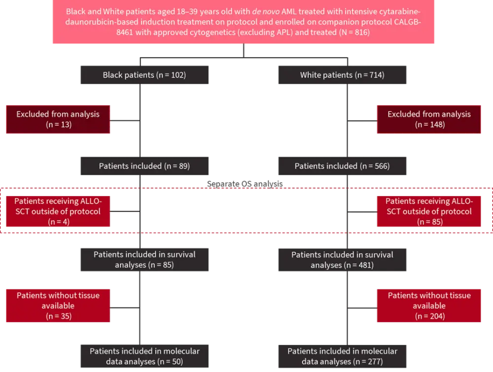
ALLO-SCT, allogeneic stem cell transplant; AML, acute myeloid leukemia; APL, acute promyelocytic leukemia; CALGB-8461, Cancer and Leukemia Group B study 8461; OS, overall survival.
*Adapted from Larkin et al.1
Results
Genetic profiling
Despite comparable clinical features at diagnosis, the cytogenetic profile of AYAs with AML differed between Black and White patients (Figure 2; Table 1). Normal cytogenetics (CN-AML) were observed in just 19% of Black patients but dominated the White patient population (40%; p < 0.001). In contrast, core binding factor AML (CBF-AML) was the most prevalent karyotype in Black AYAs with AML.
Figure 2. Genetic profiles of A Black and B White AYAs with AML*
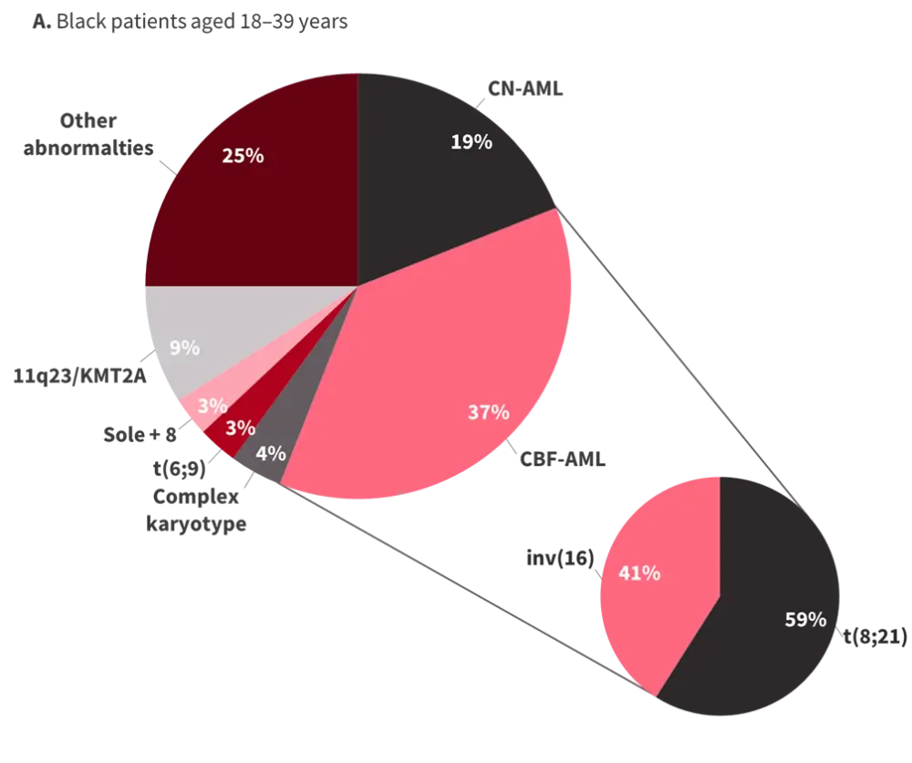
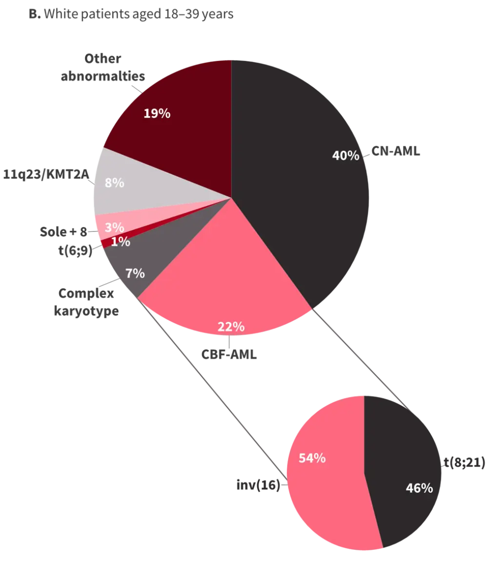
AML, acute myeloid leukemia; AYAs, adolescents and young adults; CN-AML, cytogenetically normal AML; CBF-AML, core binding factor AML.
*Adapted from Larkin et al.1
Significantly increased frequencies of CEBPA double mutant and NPM1 mutations were found in White AYAs with AML of, while mutations in ASXL1, KRAS, and ZRSR2 were more common in Black patients (Table 1).
Table 1. Mutational profile differences in AYAs with AML*
|
AML, acute myeloid leukemia; AYAs, adolescents and young adults. |
|||
|
Variant, % |
Black patients |
White patients |
p value |
|---|---|---|---|
|
Increased prevalence in Black patients |
|||
|
ASXL1 |
12 |
1 |
<0.001 |
|
BCOR |
8 |
2 |
0.05 |
|
CALR |
8 |
2 |
0.05 |
|
KRAS |
16 |
5 |
0.01 |
|
ZRSR2 |
6 |
0 |
0.01 |
|
Increased prevalence in White patients |
|||
|
CEBPA (dm) |
3 |
17 |
0.02 |
|
NPM1 |
4 |
29 |
0.01 |
Patient outcomes
Inferior outcomes were observed in Black AYAs with AML, as presented in Tables 2 and 3. A focused analysis of patients aged 18–29 years highlighted the influence of this age range on survival outcomes between Black and White patients with AML. Disparities observed in patients aged 18–29 did not persist in the 30–39 age range (Table 3). Strikingly, overall survival (OS) in Black patients aged 18–29 was 1.3 years compared with 10.2 years in White patients of the same age, indicating a survival gap of almost 10 years.
Table 2. Patient outcomes in AYAs with AML by race*
|
CR, complete remission; OS, overall survival. |
|||
|
Outcome |
Black patients |
White patients |
p value |
|---|---|---|---|
|
Early death, %† |
11 |
2 |
<0.001 |
|
CR, % |
73 |
82 |
0.06 |
|
Median OS, years |
1.5‡ |
3.1§ |
0.002 |
Table 3. Patient outcomes in AYAs with AML by age category and race*
|
CR, complete remission; DFS, disease-free survival; OS, overall survival. |
||||||
|
Outcome |
18–29 years |
30–39 years |
||||
|---|---|---|---|---|---|---|
|
Black patients |
White patients |
p value†† |
Black patients |
White patients |
p value |
|
|
Early death, % |
16 |
3 |
0.002 |
7 |
2 |
0.12 |
|
CR, % |
66 |
83 |
0.01 |
80 |
81 |
0.84 |
|
Median OS, years |
1.3 |
10.2† |
<0.001 |
2.2‡ |
2.2§ |
0.49 |
|
Median DFS, years |
1.2‖ |
1.8¶ |
0.55 |
1.2# |
1.4** |
0.55 |
CBF-AML
Due to the notable proportion of Black AYA patients presenting with CBF-AML at diagnosis, the study also sought to determine the impact of disease karyotype on patient outcome. Complete response, early death, and disease-free survival were similar between Black and White patients with CBF-AML; however, median OS was considerably poorer in Black AYA patients with CBF-AML (5.1 years vs not reached, p = 0.05).
In AYAs with AML aged 30–39 years, CBF-AML was a positive prognostic indicator across Black and White patients. However, survival outcomes in Black patients aged 18–29 years with CBF-AML, although superior to non-CBF-AML, were equivalent to those of White patients with non-CBF-AML. Furthermore, OS rates of Black patients in this age group with non-CBF-AML were particularly dismal, with a 5-year OS rate of just 12%. Taken together, these data suggest that the high proportion of Black AYAs with CBF-AML may, in fact, positively skew survival data in this patient population.
Treatment response
In a separate analysis of patients who received allogeneic stem cell transplant off protocol (Figure 1; Black, 4.5%; White, 15%), White patients exhibited significantly longer disease-free survival compared with Black patients.
Clonal patterns
Diagnosis and relapse samples were available for four Black patients, allowing paired genome profiling to evaluate the influence of genetic factors on survival in Black AYAs with AML. In all patients, dominant clones persisted from diagnosis through relapse, demonstrating clonal stability.
Conclusion
In the AML setting, patient karyotype, survival outcomes, and treatment responses vary drastically between Black and White AYA patients. Regarding the survival gap, there are a number of potential factors at play, and further research is required in the Black AYA AML population to improve patient outcomes.
References
Please indicate your level of agreement with the following statements:
The content was clear and easy to understand
The content addressed the learning objectives
The content was relevant to my practice
I will change my clinical practice as a result of this content
Your opinion matters
Which AML-related topic do you currently need the most practical guidance on?